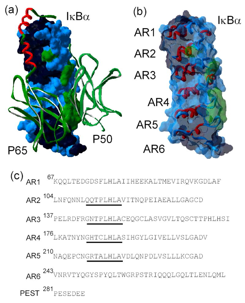
Figure 1.

a) The x-ray crystal structure of IκBα (surface representation) in complex with NF-κB (ribbon representation) (pdb accession number 1NFI) 7. The dimerization domains of NF-κB p50 and p65 are shown in green and the NF-κB p65-NLS polypeptide in red. The surface of IκBα is colored according to the NF-κB contact regions: p65 contacting residues in black, p50 contacting residues in green, non-contacting residues in blue. b) The high resolution structure of only IκBα from the structure of the complex in the same orientation as in a), with a ribbon representation of the ankyrin repeats (AR), and the same coloring scheme as in a) for the translucent surface. c) Amino acid sequence of the ankyrin repeat domain of IκBα(67 to 287), aligned according to each ankyrin repeat. The ‘consensus’ GxTPLHLA motif is underlined.
