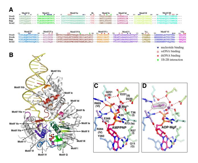
Figure 2.
The conserved sequence motifs among UvrD, PcrA, Rep and Srs2. (A) Sequence alignment of the 8 ATPase motifs (in different shades of green (domain 1A) and blue and purple (2A), and 8 newly found DNA binding and domain interaction motifs (in brown to red colors). (B) The 16 motifs are mapped onto the UvrD-DNA-AMPPNP structure using the same color scheme as in (A). (C) and (D) Coordination of the AMPPNP and ADP·MgF3 by the 8 helicase motifs. Carbon atoms of the UvrD side chains are colored light green (domain 1A) and light blue and purple (2A). Nitrogen atoms are shown in blue, oxygen in red, and Mg2+ in dark purple. Water molecules are shown as red spheres. The Fo-Fc electron density of MgF3 is superimposed onto the model.

