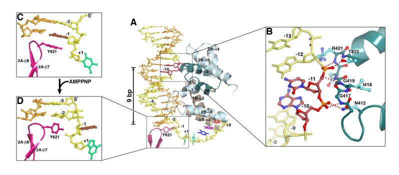
Figure 4.
Interactions between UvrD and dsDNA. (A) The four HLH structures from domains 2B (light and dark cyan) and 1B (brown) interact with 14 to 16 bps. These interactions are similar between the binary and ternary complexes, while the ternary complex structure is shown. The separation pin (Y621) buttresses the end of DNA duplex. (B) A close-up view of the GIG motif and dsDNA interactions. (C) Stacking of Y621 with the-1 bp in the binary complex. (D) Unwinding of the-1 bp in the ternary complex, and the accompanying side chain conformational change of Y621.

