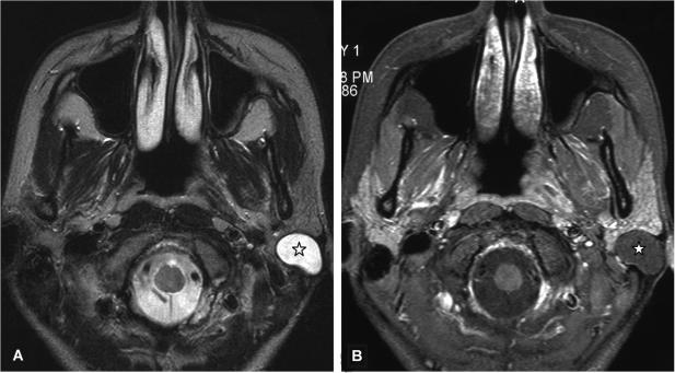
Figure 8.
Axial T2-weighted MR image (A) of a 25-year-old woman with an epidermoid cyst in the superficial lobe (asterisk) presented as a well-delineated and homogeneous hyperintense lesion. The T1-weighted contrast-enhanced fat suppressed MR image (B) at the same level shows a homogeneous hypointense lesion without contrast medium enhancement and well-delineated margins.

