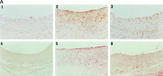
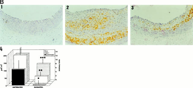
Figure 1.
MCP-1, IL-8, and macrophage detection by immunohistochemistry in arterial sections. Arterial sections were stained with anti-MCP-1, anti-IL-8, or RAM11 antibodies. A: In the upper part is shown MCP-1 immunostaining of a control (1), an untreated (2), and a quinapril-treated (3) animal. In the lower part is shown the staining for IL-8 of a control (2), an untreated (5), and a treated (6) animal. B: Staining for macrophages corresponding to a control (1), an untreated (2), and a quinapril-treated (3). Magnification, ×400. In the fourth panel of fig 1B ▶ is shown the bar graph corresponding to the mean neointimal area occupied by the macrophages (closed bars), the MCP-1 (open bars) and the semiquantitative score for IL-8 (hatched bars). The figure illustrates the association between the three parameters. *, P < 0.04; **, P < 0.05; ***, P < 0.04.


