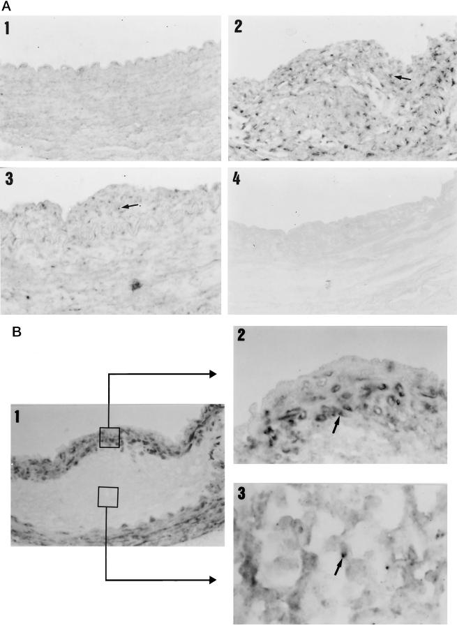
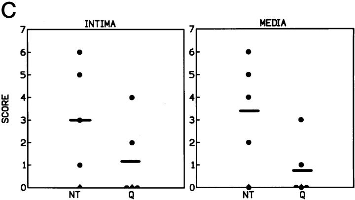
Figure 6.
Arterial localization of the NF-κB activity determined by Southwestern histochemistry. Arterial sections were hybridized with an oligonucleotide containing the consensus sequence of the NF-κB recognition site. A: Control animals (1) did not present activated nuclei, whereas untreated animals (2) showed extensive staining both in the intima and in the media (arrow). Quinapril-treated rabbits (3) showed a marked decrease in the presence of activated nuclei in both the intima and the media. Incubation of arterial sections from untreated animals with a mutant NF-κB oligonucleotide showed no staining (4). Magnification, ×400. B: In panel B 1, it can be seen a detail of a lesion showing the presence of VSMC in brown and macrophages in light pink, (this section corresponds to the untreated one shown in panel A2). Magnification, ×400. A detail of the localization of the NF-κB activity by double staining with anti-α actin antibody is shown in B2 and with -macrophage in B3 (Magnification, ×1000). The arrows indicate the presence of a nuclei stained for NF-κB and surrounded by a cytoplasm stained with the antibody specific for VSMC (B2) and macrophages (B3). C: The graph shows the semiquantitative score of the NF-κB activity in the intima and media of the untreated (NT) and quinapril-treated (Q) animals.


