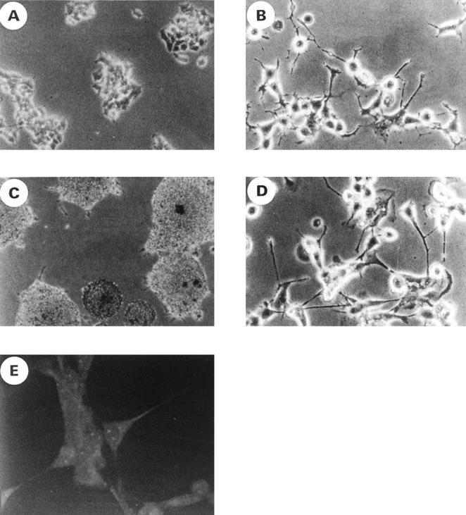
Figure 2.

The morphological changes and neurofilament expression after the differentiation induction of ES cells with db-cAMP. A–D: Cell culture morphology of RD-ES (A and B) and CADO-ES1 (C and D) cells under phase contrast microscopy. RD-ES and CADO-ES1 cells were cultured in the absence (A and C) or presence (B and D) of db-cAMP for 12 days. The differentiated cells show long cytoplasmic processes, and some neurites terminated in growth cones (B and D). E: Differentiated CADO-ES1 cells were stained with anti-NF-H MAb and analyzed by confocal microscopy. Note the neuritic process and perinuclear localization of NF-H.
