
Figure 3.
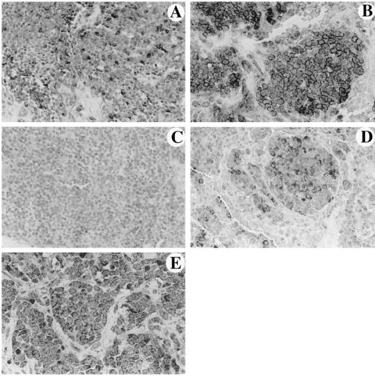
Primary (A) (Magnification, ×240) and metastatic (B) (Magnification, ×450) tumor specimens from different patients stained for FasL. Primary (C) (Magnification, ×165) and metastatic (D) (Magnification, ×250) tumor specimens from the same patient stained for FasL. FasL-positive cells exhibit diffuse cytoplasmic and/or peripheral membrane staining in all positive cases. The number of positive cells is higher in the metastatic tumors shown in B and D. In the paired specimens C and D, there is a definite increase in the number and immunoreactivity of FasL-expressing cells. E: ESFT cells exhibit diffuse cytoplasmic staining for Fas (Magnification, ×520).