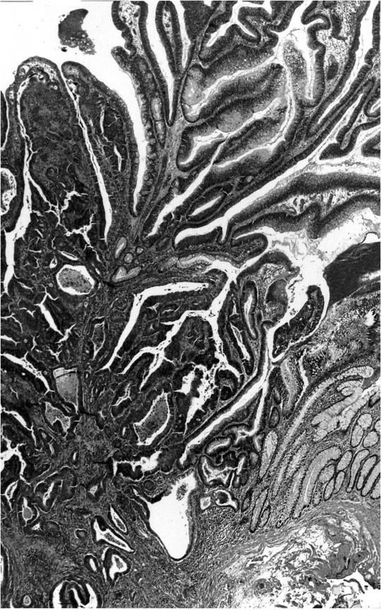
Figure 2.

Example of an ex-adenoma carcinoma. This lesion was a polypoid adenoma 1.5 cm in diameter and contained an area of carcinoma invading the submucosa seen in the left hand side of the picture. Remaining adenoma elements are seen in the upper right and nonneoplastic-appearing mucosa in the lower right (H&E stain, original magnification, ×25).
