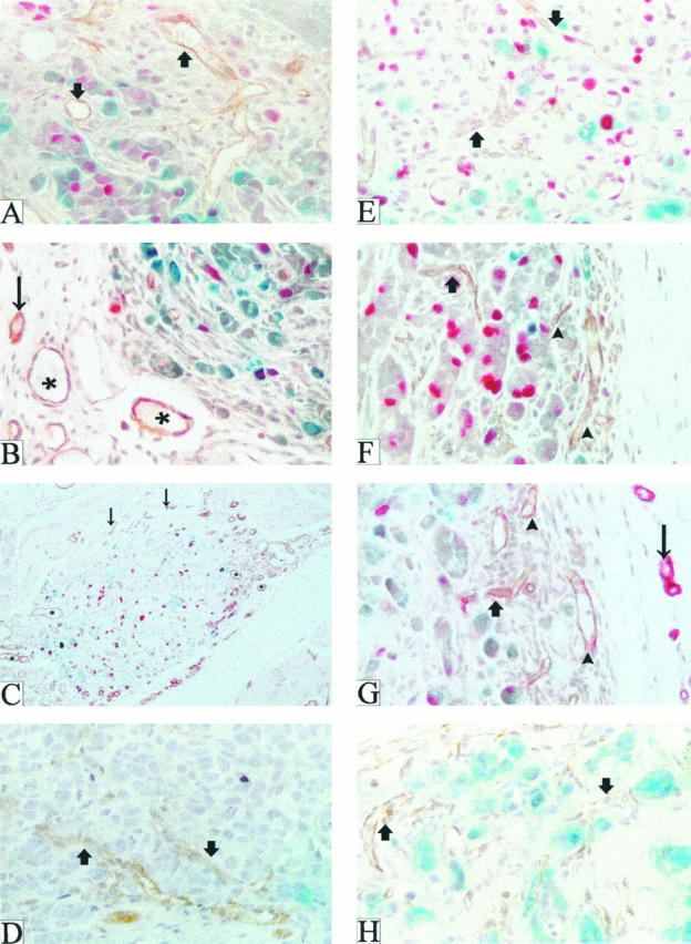
Figure 1.

Patterns of neovascularization after injection of parental MCF-7 and FGF-transfected MCF-7 cells into the mammary fat pads of ovariectomized nude mice. Tumors or tumor nodules produced by injection of parental MCF-7 (A–C) or FGF-4 transfected (MKL-F, E–G) cells were harvested at days 6 (A and E), 15 (B and F), and 35 (C and G), stained with X-gal (blue) to reveal tumor margins, embedded in paraffin, sectioned, and subjected to double immunohistochemistry for BrdU (red) and murine PECAM-1 (brown or reddish brown). (Because the tumors were stained with X-gal when whole, only surface tumor cells stain blue.) D and H depict VEGF/VPF-transfected cell tumors (MV165-14) harvested 18 days after tumor cell injection. Arrowheads point to edge-associated microvessels, thick arrows to intratumor microvessels; asterisks denote ectatic stromal vessels, and thin arrows indicate normal stromal vessels. Magnification, ×400 (A, B, and D–H) and ×200 (C).
