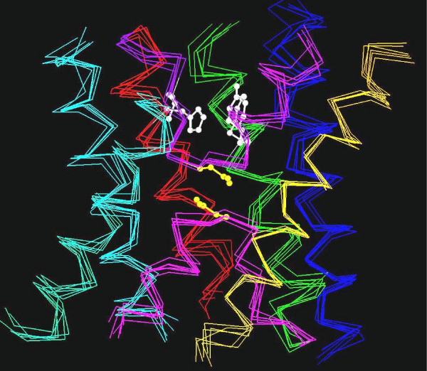
Figure 1.

Superposition of aquaporin crystal structures. The transmembrane regions of six aquaporin crystal structures (bovine AQP1, E. coli AqpZ, sheep AQP0, spinach plasma membrane aquaporin SoPIP2;1, archaeal aquaporin AqpM from Methanothermobacter marburgensis and E. coli GlpF) are superposed. The corresponding PDB IDs are 1J4N, 1RC2 (B chain), 2B6O, 1Z98 (A chain), 2F2B and 1FX8 respectively. For clarity, Cα traces of only the six transmembrane helices and the loops B and E are shown: TM1 – blue, TM2 – green, loop B – pink, TM3 – orange, TM4 – red, loop E – purple, TM5 – cyan and TM6 – green. The residues forming the Ar/R selectivity filter from SoPIP2;1 are shown in white and the asparagines from the conserved NPA motif of loops B and E are shown in yellow. The aquaporin structures from bacteria, archaea, plant and mammals show a conserved "hour-glass" fold and the helices form a right-handed bundle structure.
