
Figure 1.
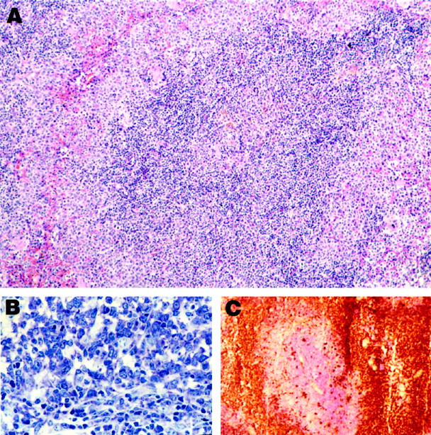
Histology and immunocytochemistry of MZL. A: MZL in low magnification. Note that the cells at the borders (marginal zones) are somewhat larger and more lightly stained than those in the center. H&E; magnification, ×110. B: Higher magnification of MZL, showing the high degree of basophilia in MZL cells. Giemsa; magnification, ×200. C: Frozen section, showing staining of MZL cells for IgM. The central portion, counterstained with hematoxylin, represents the T cells of the PALS. magnification, ×50.