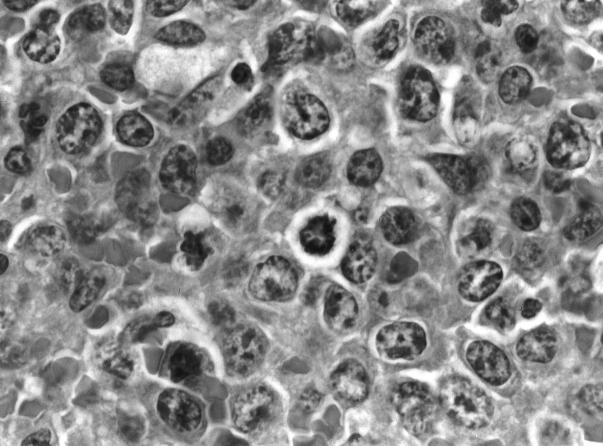
Figure 2.
Typical MZL++ composed of cells with CB-like morphology and with numerous mitotic figures. A biopsy taken from the same spleen 113 days earlier was diagnosed as EMZ because the marginal zone was only slightly enlarged and was composed of typical marginal zone cells. The biopsy had three IgH rearrangements, but only one remained at necropsy.

