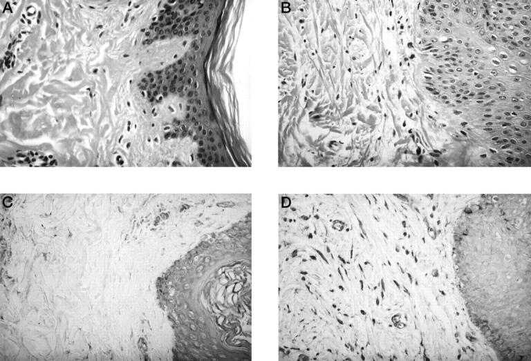
Figure 2.

Histological comparison between normal and keloid tissues. A: H&E-stained sections of normal tissues (N-1). B: H&E-stained sections of keloid tissues (K-1). The keloid contains numerous blood vessels, collagen fibers, and fibroblasts. C: Immunohistochemistry of normal tissues (N-1) for IGF-IRβ subunits. Note the strong staining in the epidermis and endothelial cells and weak staining in the fibroblasts. D: Immunohistochemistry of keloid tissues (K-1) for IGF-IRβ subunits. Note the presence of strongly stained cells in the epidermis, endothelial cells, and fibroblasts. Magnification, ×400.
