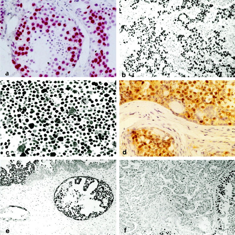
Figure 1.

a: Section of a normal adult testis stained with Ki-A10; spermatogonia are consistently positive (red label); the staining intensity decreases in spermatocytes along with maturation and becomes undetectable in spermatids and spermatozoa. Sertoli cells show no reactivity (original magnification, ×350). b: Strongly Ki-A10-labeled tumor cell nuclei of testicular seminoma scattered in a lymphoid infiltrate negative to Ki-A10 (magnification, ×140). c: The totality of the bizarre cells of spermatocytic seminoma exhibit strong nuclear Ki-A10 reactivity (magnification, ×350). d: The germ cell component of gonadoblastoma stains with slightly variable intensity (brown label) for Ki-A10 (magnification, ×140). e: A large number of cells in YST of glandular type express the Ki-A10 antigen; focally, abrupt loss of antigenity is noted (upper right) (magnification, ×140). f: Embryonal carcinoma characteristically does not express the Ki-A10 antigen; in the preserved seminiferous tubules (bottom right), spermatogonia and spermatocytes are strongly positive (magnification, ×140; all sections peroxidase technique and hematoxylin counterstain).
