
Figure 4.
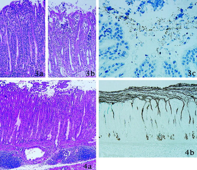
8 weeks after inoculation of H. pylori. a: Erosion is observed. Small clusters of surface mucous cells protruded into the lumen. Dilated mucous gland and accumulation of lymphocytes are present in the submucosa. b: Mucins in the surface mucous cells and in the pyloric glands are depleted markedly. Surface mucous gel layer (SMGL) was thickened and consist of two type of gastric mucins. Thin strands of mucins from pyloric gland cells are evident and join the SMGL (GOTS/PCS).
Figure 3. 4 weeks after inoculation of H. pylori. a: In the transitional zone between pylorus and fundus, elongated pseudopyloric glands emerge (H&E stain). b: Neutrophils infiltrate the epithelium and form intrafoveolar microabscess (H&E stain). c: H. pylori stained brown with immunostaining for H. pylori and are present in the surface mucous gel layer (immunoperoxidase method for H. pylori).