
Figure 8.
52 weeks after inoculation of H. pylori. a: Dilated mucous glands are distributed in the lamina propria and submucosa (H&E stain). b: These mucous glands stain brown with PCS (GOTS/PCS).
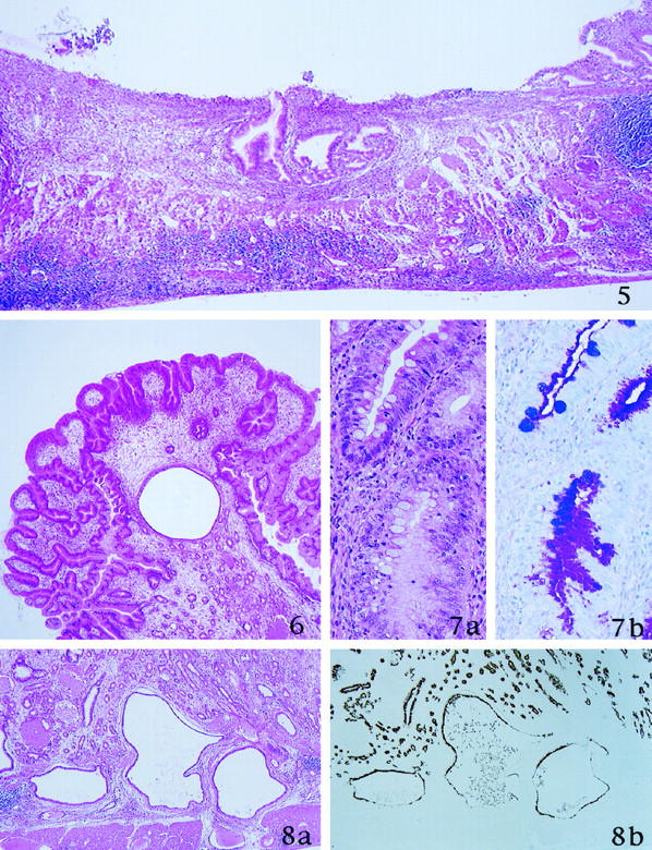
Figure 5. 26 weeks after inoculation of H. pylori. Ulcer is formed. Granulation tissue with inflammatory cells and necrotic materials are present in the ulcer base (H&E stain).
Figure 6. 26 weeks after inoculation of H. pylori. Histological finding of the sessile polyps around the border between pylorus and fundus. Hyperplastic foveolae with widened stromal tissue are evident (H&E stain).
Figure 7. 26 weeks after inoculation of H. pylori. a: Incomplete intestinal metaplasia is recognized. b: Metaplastic goblet cells show alcianophilia (AB/PAS stain).