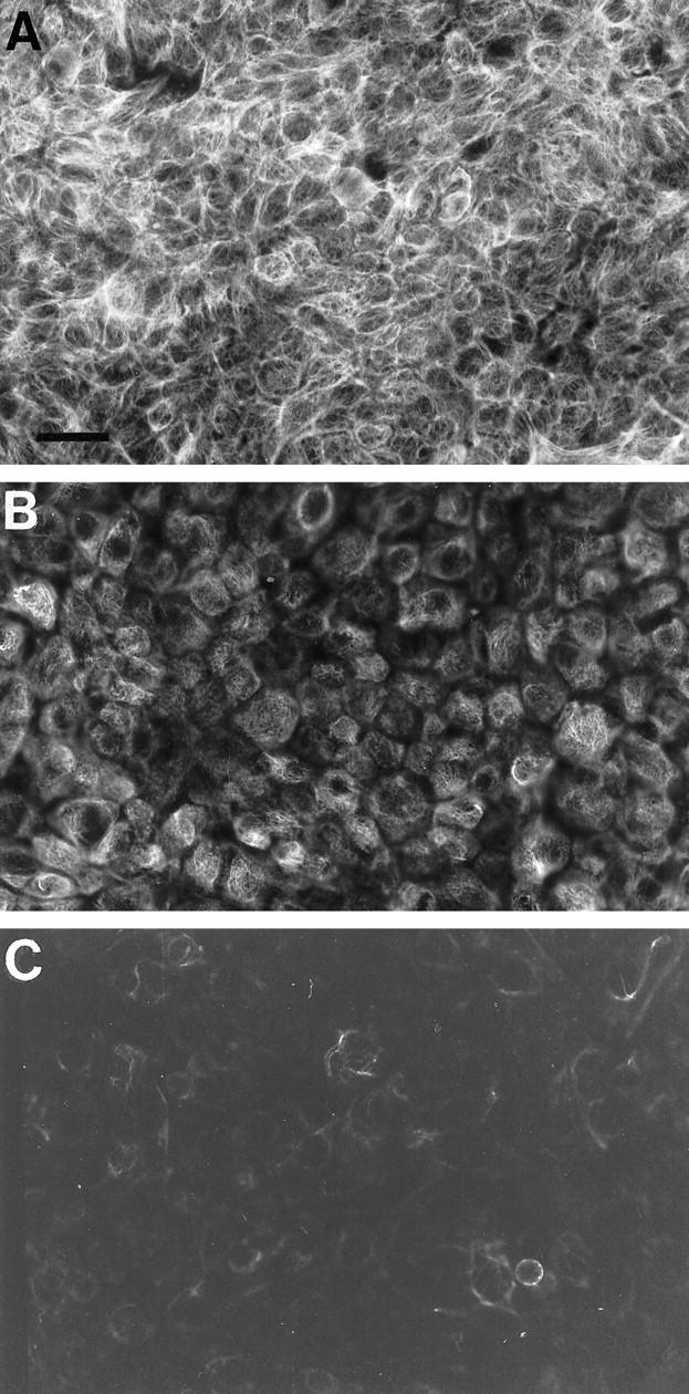
Figure 3.

Indirect immunofluorescence labeling of cultured urinary bladder cancer cells with neurofibromin-specific antibody. Representative areas of each specimen were photographed using the same exposure times, and the photomicrographs were reproduced under identical conditions. A: RT4 cells originating from grade 1 urinary bladder carcinoma. B: 5637 grade 2 cancer cells. C: T24 grade 3 cancer cells. The intensity of the immunosignals was quantitated using digital image analysis system MCID-M4 (see Results). Magnification, ×40; scale bar, 25 μm.
