
Figure 1.
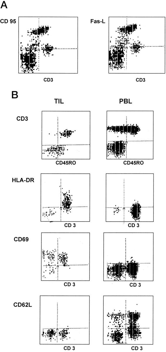
Lymphocytes infiltrating colorectal hepatic metastases comprise mainly activated memory T cells expressing Fas (CD95) and Fas-L. A: Phenotype of freshly isolated TIL showing that they include a subset of activated lymphocytes that are CD95high and Fas-Lhigh. B: The majority of the TIL are CD45RO+, CD69high, HLA-DRhigh, and CD62L (L-selectin)low compared with autologous peripheral blood lymphocytes (PBL). Cells were analyzed by flow cytometry gated on CD3+ population. Figure ▶ shows one representative experiment of ten.