
Figure 4.
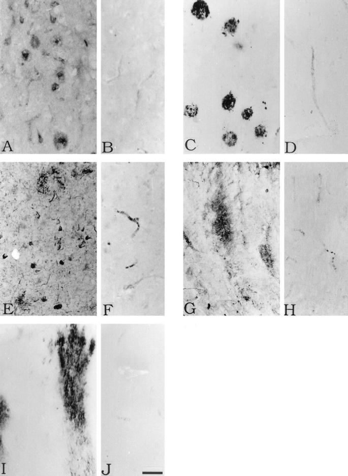
Immunohistochemical staining of AD transentorhinal cortex (A, C, E) and cerebellum (G, I) and control transentorhinal cortex (B, D, F) and cerebellum (H, J). Staining is for C1q (A, B, G, H), C4d (C, D, I, J) and C5b-9 (E, F ). See under Methods for details. In A, the C1q antibody stains senile plaques as well as tangled neurons very weakly. In G, the C1q antibody stains diffuse amyloid deposits. In C, the C4d antibody stains senile plaques, and in I it stains diffuse amyloid deposits. In E, the anti-C5b-9 antibody stains dystrophic neurites and tangled neurons. The control sections B, D, F, H, and J show no specific staining of brain structures. Scale bar in J = 50 μm.