
Figure 2.
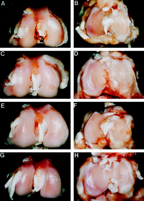
Macroscopic appearance of cartilage from femoral condyles (left panels) and tibial plateaus (right panels) of (A,B) a 4-week OA rabbit showing pitting of the central, weight-bearing region of the medial condyles and plateaus; (C,D) an 8-week OA rabbit injected with the control plasmid showing erosion and pitting of the medial condyle and plateau; (E,F) an 8-week OA rabbit treated with 500 μg of IL-1Ra plasmid showing pitted areas of cartilage on medial condyle and plateau; (G,H) an 8-week rabbit treated with 1000 μg of IL-1Ra plasmid showing pitted areas of cartilage on the medial condyle and plateau resembling those of a 4-week OA rabbit.