
Figure 3.
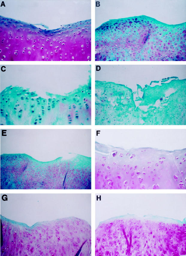
Representative sections of articular cartilage from a medial femoral condyle (left panels) and tibial plateau (right panels) of (A,B) a 4-week OA rabbit; (C,D) an 8-week OA rabbit injected with the control plasmid; (E,F) an 8-week OA rabbit treated with 500 μg IL-1Ra plasmid; (G,H) an 8-week rabbit treated with 1000 μg IL-1Ra plasmid. safranin-O staining. Original magnification, ×250 (A, C, F), ×100 (B, D, E, G, H).