
Figure 2.
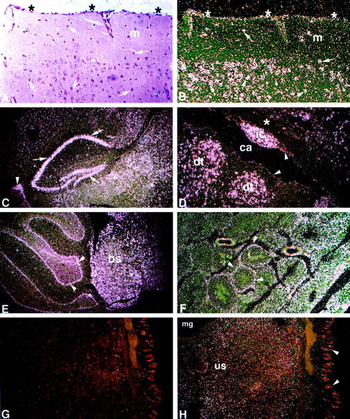
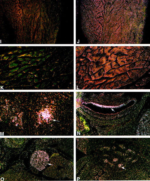
Spatial and temporal expression of the mouse GAA mRNA by in situ hybridization. A: Bright-field image with H&E. B to K: Dark-field images for the sense (G, I, and K) and for the antisense (B–F, H, J, and L–P) 35S-labeled riboprobes. A to L: Adult tissues. High-intensity signal is present in neurons throughout the neuraxis (B–E). A and B: Cerebral cortex. Pyramidal and granule neurons (arrows) have large purple nuclei by H&E (A) and show high-intensity signal (B). Signal of lower intensity is in the molecular layer (m). Moderate-intensity signal is in meningeal cells of the pia mater-arachnoid (*). C: Cerebral sagittal section. Highest-intensity signals are in neurons of the hippocampus (arrows), cells lining the choroid plexus (arrowhead), and surrounding neuronal layers. D: Cerebrum/aqueduct of Sylvius. High-intensity signal is in neurons adjacent to the cerebral aqueduct (ca) located in the inferior colliculus nucleus (ic) and dorsal tegmental nuclei (dt). Low- to very-low-intensity signal is in glial cells of the white matter (*) and ependymal lining cells (arrowheads). E: Hindbrain. High-intensity signal is in neurons of the cerebellar Purkinje cell layer (arrowheads) and neurons of the brainstem (bs). F: Testis. Signal of moderate intensity is in seminiferous tubules (arrowheads). G and H: Uterus. Uterine stromal mucosal cells (us), negative with the sense riboprobe (G), show diffuse low signal with the antisense riboprobe (H). Very low signal is in mucosal glandular (mg) and epithelial cells (arrowheads). I and J: Heart/ventricle. Diffuse signal of moderate intensity relative to background levels in the sense control (I) is in cardiomyocytes (J). K and L: Paraspinal muscle. Low level signal above background levels (K) is in myocytes (L). M to P: Embryonic tissues, 16 day. M: Hypothalamus. Moderate level signal is in differentiating neurons (arrow). N: Inner ear. Moderate level signal is in the epithelial lining cells (arrow). O: Sympathetic ganglia. Moderate signal is in neurons (arrow). P: Testis. Moderate signal is in epithelial cells of the seminiferous tubules (arrow). Counterstaining is with H&E. Magnification, ×200 (A and B), ×100 (D and F–P), and ×40 (C and E).