
Figure 1.
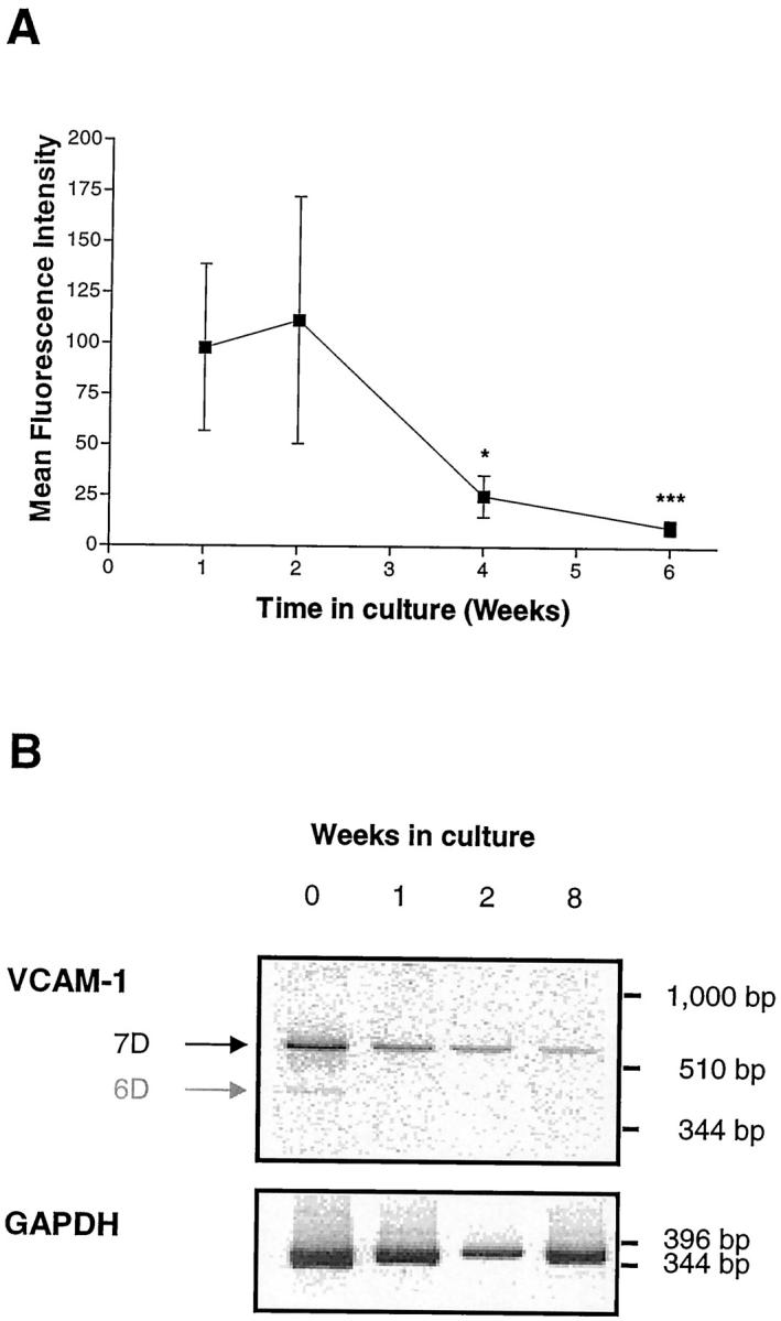
Elevated expression of VCAM-1 cell surface protein (A) and mRNA (B) by freshly isolated rheumatoid fibroblast-like synoviocytes declines with increasing time in culture. FLSs were isolated as described in Methods and cultured with 10% FBS for various periods of time. Medium was replenished every 5 days. A: cell surface VCAM-1 expression was analyzed after 1, 2, 4, and 6 weeks by flow cytometry using mAb BBA-5. Samples were analyzed using a FACScan flow cytometer and the results expressed as corrected mean fluorescence intensity. Data are presented as the mean ± SE of four experiments. * indicates p < 0.05, and *** p < 0.001 (compared with the results at week 1). B: VCAM-1 mRNA levels after 0 (ie, from freshly isolated synovial cells), 1, 2, and 8 weeks. Total RNA was isolated and semiquantitative RT-PCR performed using VCAM-1-specific and GAPDH-specific primers (see Methods). PCR products were visualized after electrophoresis on an 8% polyacrylamide gel and exposure to a phosphorimager. Note the presence of both the 6D and 7D mRNA transcripts in freshly isolated synovial cells.