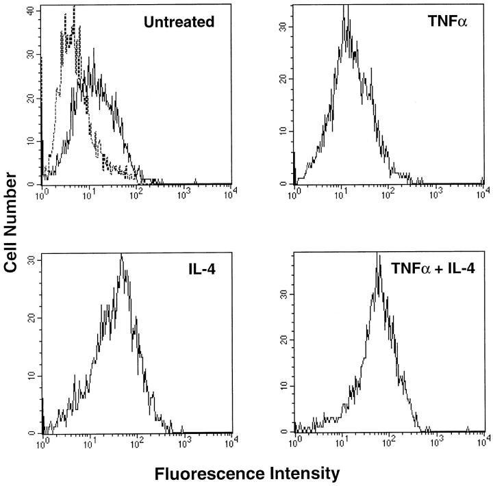
Figure 4.
Flow-cytometric analysis of VCAM-1 on RA fibroblast-like synoviocytes treated with TNF-α and IL-4. Cells were cultured with either medium only (untreated), TNF-α (10 ng/ml), IL-4 (10 ng/ml), or TNF-α plus IL-4 for 3 days. Flow cytometry for cell surface VCAM-1 was carried out using mAb BBA-5 as primary antibody. Samples were analyzed using a FACScan flow cytometer. The dashed histogram (in untreated cells) represents staining with PE-conjugated anti-mouse IgG only. Solid line histograms represent cells stained with anti-VCAM-1 and PE-conjugated anti-mouse IgG. Results shown are representative of four experiments.

