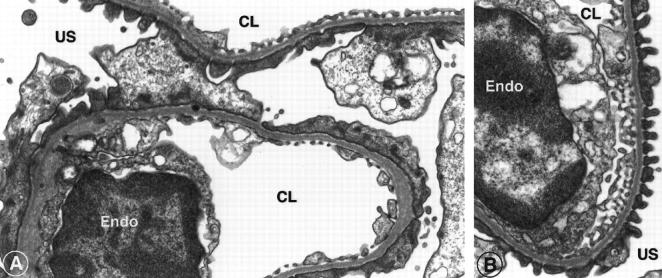
Figure 5.
Comparison of foot process architecture by electron microscopy of PBS-infused control Mpv17−/− mice (A), and of animals infused with DMTU via an implanted minipump for 14 days (B). Tissue samples were collected on day 14 of the infusion from the subcortical area. Untreated Mpv17−/− mice show extensive flattening of the foot processes, whereas the DMTU-treated group consistently showed normal podocyte foot processes and slit diaphragms. E, endothelium; CL, capillary lumen; US, urinary space. Original magnification, ×28,000.

