
Figure 4.
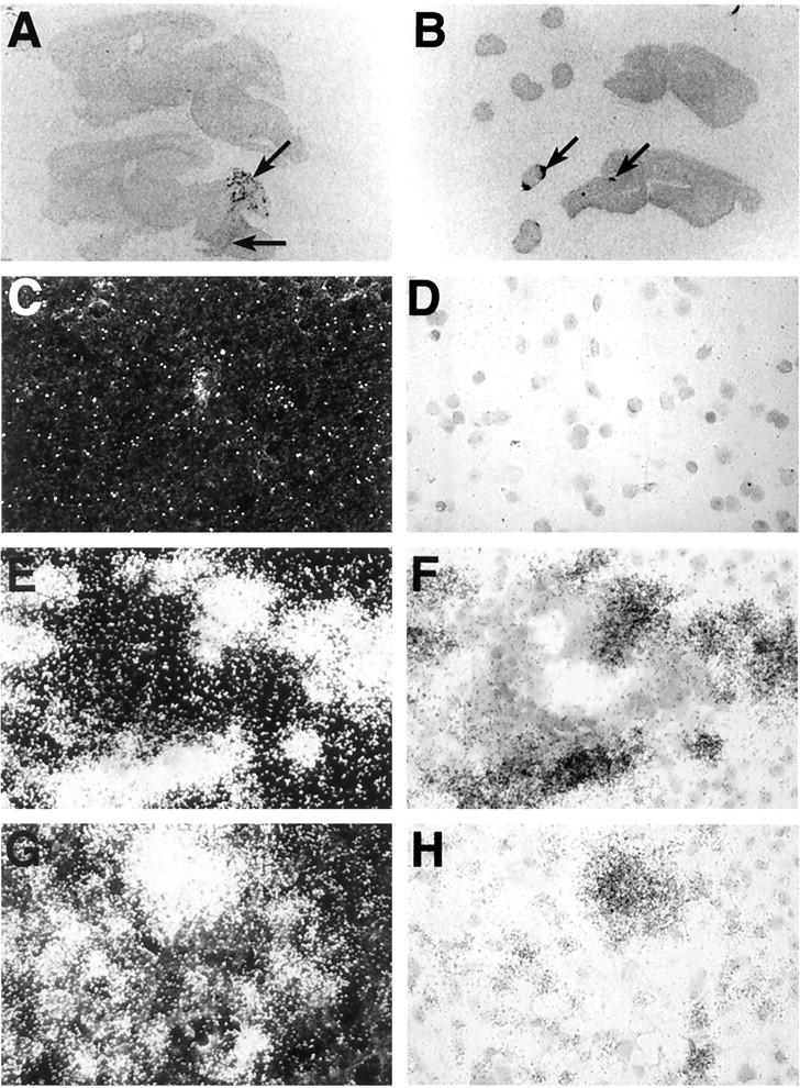
Anatomical and microanatomical localization of C10 RNA in brain from GFAP-IL3 mice or brain and spinal cord from mice with MBP-EAE. Images from Cronex film (5 day exposure) of sagittal sections (10 μm) from GFAP-IL3 transgenic mice (A) or mice with EAE (B). The sections located at the top in A and B represent tissue from wild-type control mice. Sections were hybridized with 35S-labeled antisense RNA probe to C10 RNA as outlined in Materials and Methods. Areas of probe hybridization are indicated by the arrows. For microanatomical analysis, sections hybridized with the 35S-labeled antisense probe to C10 RNA were coated with photographic emulsion, developed after 1 week, and visualized using dark-field (C, E, G) or bright-field (D, F, H) microscopy. No hybridization was detected in brain or spinal cord from control mice (C, D). In cerebellum from GFAP-IL3 mice (E, F), intense hybridization signal is shown surrounding a vascular lymphocytic cuff. In EAE (G, H), hybridization signal was closely associated with the infiltrating mononuclear cell population in the spinal cord.