
Figure 1.
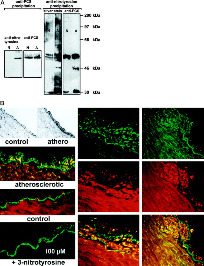
Nitrated protein and PCS in atherosclerotic bovine coronary arteries. A: Protein from normal (N) or atherosclerotic (A) bovine coronary arteries was immunoprecipitated either with an anti-PCS (left) or an anti-nitrotyrosine (right) antibody. Protein precipitated by anti-PCS was separated electrophoretically and immunostained with anti-nitrotyrosine or anti-PCS antibodies. Proteins precipitated by anti-nitrotyrosine were separated similarly and then either silver stained or analyzed by immunoblot with anti-PCS antibodies. Molecular mass of standard proteins run on the same gel are indicated. B: Top left, representative examples for normal (control) or early atherosclerotic vessels (athero) used in this study. Dark color in the thickened athero endothelium is due to CD11b immunoreactivity (macrophage marker); middle left, intimal thickening in atherosclerotic vessel that is strongly immunopositive for nitrotyrosine (green). PCS is stained in the intima and media (red). Below, control tissue is shown where nitrotyrosine staining is very weak and the endothelium is undisturbed. Bottom left, an intimal thickening was stained for nitrotyrosine in the presence of 10 mmol/L free 3-nitrotyrosine. Only the autofluorescence of the basal membrane is visible. Middle and right columns , more advanced lesions (top-green, protein-nitrotyrosine; middle-red, PCS; bottom, overlay image with yellow indicating colocalization). Nitrotyrosine is detectable also in the subintimal media below the strongly developed lesion shown to the right.