
Figure 2.
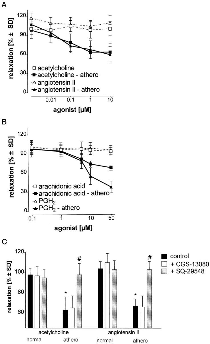
Defective relaxation of atherosclerotic aortic strips due to impaired PCS activity. A and B: Aortic strips were exposed to different concentrations of contractile stimuli. The extent of subsequent relaxation to the original length was quantitated in control tissue (open symbols) and atherosclerotic tissue (filled symbols). C: In some experiments, aortic strips were preincubated with CGS-13080 (10 μmol/L) or SQ-29548 (10 μmol/L) before stimulation with acetylcholine (10 μmol/L) or angiotensin II (10 μmol/L). Data represent means ± SD from five experiments. * P < 0.01 (atherosclerotic versus normal tissue); # P < 0.01 (SQ-29548 versus atherosclerotic).