
Figure 4.
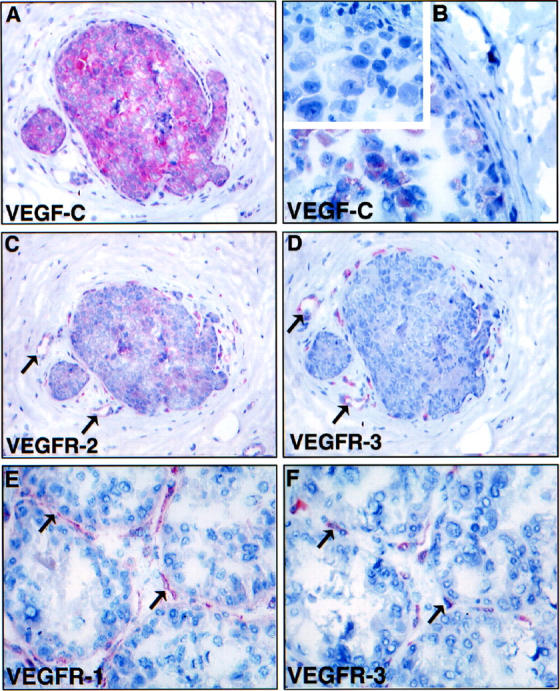
Comparison of VEGF-C (A, B) and VEGFR (C-F) stainings in intraductal carcinoma. Note the expression of VEGF-C in all (A) or part (B) of the carcinoma in situ cells. Negative control using anti-VEGF-C antibodies blocked with a 10-fold molar excess of the VEGF-C protein gave no staining of the carcinoma cells (inset in B). C and D show sections adjacent to A stained for VEGFR-2 and VEGFR-3, respectively. Note that the necklace vessels are positive for both VEGFR-2 and VEGFR-3 in the vicinity of VEGF-C expressing carcinoma cells. E and F show a similar comparison for VEGFR-1 and VEGFR-3, respectively. Note that the staining for VEGFR-3 appears more discontinuous. Magnification, ×300 (<label;A, C, D>); ×400 (B); ×350 (E, F).