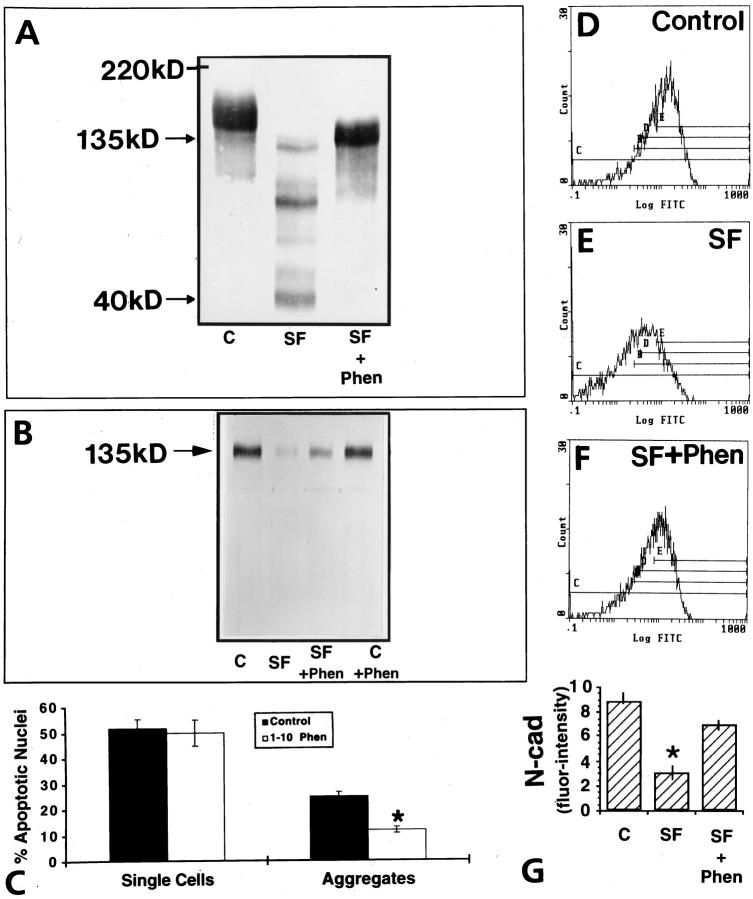
Figure 10.
Effect of 1–10 phenanthroline on N-cadherin expression and apoptosis on GCs in culture. Immunoblotting for N-cadherin by utilizing the 13A9 antibody against the cytoplasmic domain of N-cadherin (A) or the GC4 antibody against the extracellular domain of N-cadherin (B). Note the reduction of the 135-kd N-cadherin protein in GCs in the absence of serum (A and B, SF) and the appearance of lower molecular weight fragments when the cytoplasmic domain antibody was used (A). In contrast, 1–10 phenanthroline reverses this effect (A and B). C: Note that the metalloproteinase inhibitor had no effect on the percentage of apoptotic single cells but significantly decreased (*P < 0.05) the percentage of apoptotic nuclei in cellular aggregates. D to F: Flow cytometric analysis for N-cadherin of one representative experiment showing the clear shift to the right when cells were treated for 24 hour with 3 mmol/L 1–10 phenanthroline (NS+Phen). G: Quantitative analysis clearly indicates a significant increase of immunodetectable N-cadherin in the presence of 1–10 phenanthroline (*P < 0.05).

