
Figure 6.
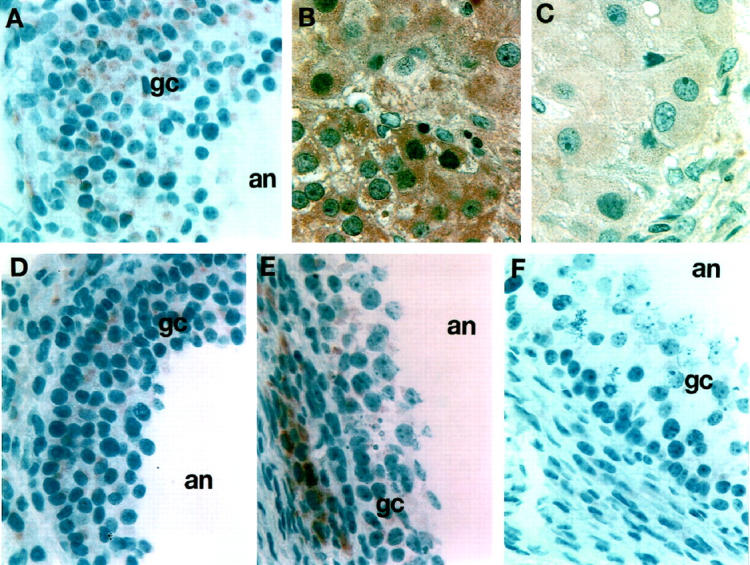
Immunohistochemical detection of N-cadherin in adult human ovary in situ. A: Normal antral follicle with multiple layers of GCs (gc) shows N-cadherin staining through all layers extending to the follicular antrum (an). B and C: Extensive staining for N-cadherin is observed in luteal cells and is more marked in the early/mid luteal phase (B) and is much weaker in a corpus luteum obtained from the late luteal phase (C). D to F: N-cadherin immunostaining of atretic ovarian follicles (at progressive stages early to late D through F), which clearly show limited numbers of GCs that appear to be losing their cell-cell junctions and their cellular integrity. Note the limited staining for N-cadherin of the granulosa cells, which is either almost absent or localized to cells closest to the basement membrane. Magnification, ×300.