
Figure 3.
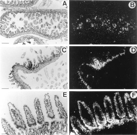
In situ hybridization of 35S-labeled antisense mouse HIP/PAP probe to mouse intestine sections under bright-field (A,C,E) and dark field (B,D,F) illumination. A and B: Longitudinal section through small intestine of E16.5 mouse embryo. C and D: Section of mouse newborn intestine. E and F: Section of mouse adult jejunum. Scale bar, 250 μm (A,B,C,D); 100 μm (E,F).