
Figure 4.
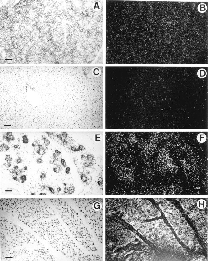
In situ hybridization of 35S-labeled antisense mouse HIP/PAP probe to sections of liver and pancreas under bright field (A,C,E,G) and dark field (B,D,F,H) illumination. A and B: Section through liver of a E16.5 mouse embryo; C and D: Section of mouse adult liver; E and F: Section through pancreas of a E16.5 mouse embryo; G and H: Section of mouse adult pancreas (10-day exposure time). Note that the white specks seen in B are caused by the refringent membrane of erythrocytes and not to silver grains. Scale bar, 250 μm (A,B,C,D); 100 μm (E,F,G,H).