
Figure 5.
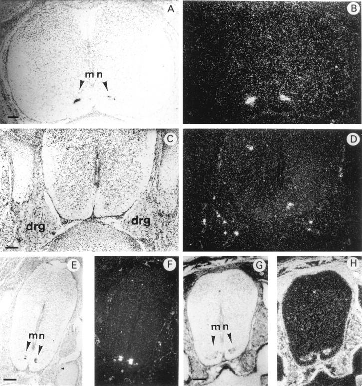
In situ hybridization of 35S-labeled antisense mouse HIP/PAP or mouse Galectin 1 probe to transverse sections through the spinal cord of E16.5 mouse embryo under bright field (A,C,E,G) and dark field (B,D,F,H) illumination. A and B: cervical region; C–H: lumbar region. For A–F the probe was mouse HIP/PAP, for G and H the probe was Galectin 1 cDNA (scale bars, 250 μm). mn, motor neuron. Note dorsal root ganglia (DRG) in C and D. DRG are aggregate of neuronal cell bodies that transmit somatic sensory information from periphery to spinal cord. Dorsal side towards the top, ventral side towards the bottom of the photographs.