
Figure 1.
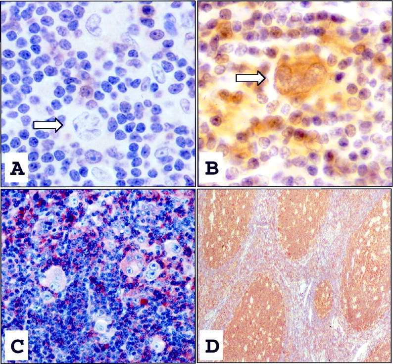
This L&H cell (arrow), as seen in the majority of NLPHD cases, was immunohistochemically negative for caspase-3, whereas scattered plasma cells and lymphocytes expressed caspase-3 (A, DAB ×1000). A single L&H cell (arrow) from one case of NLPHD displayed cytoplasmic expression of caspase-3 (B, DAB ×1000). Control cases of nodular sclerosis Hodgkin’s disease demonstrated diffuse caspase-3-immunopositivity of HRS cells and intense positive immunostaining of lymphocytes and plasma cells within the surrounding infiltrate (C, DAB ×400). Reactive follicular centers in tonsil controls also displayed intense positive staining for caspase-3 (D, DAB, ×200).