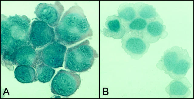
Figure 2.
Immunohistochemical detection of caspase-3 on cytospin preparations of the L428 cell line (A) displayed strong cytoplasmic positive staining for caspase-3 (AEC, ×400); however, the KMH2 cell line (B) failed to express caspase-3 (AEC, ×400). Isotype control antibody staining was negative (data not shown).

