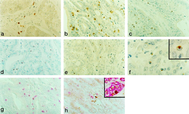
Figure 1.
Characterization of intraepithelial lymphocytes in representative cases of MSI− (a) and MSI+ (b to g) CRCs. Activated CTLs infiltrating within neoplastic epithelial structures are present in significantly higher numbers in MSI+ CRCs. CD8 (a and b), TIA-1 (c), perforin (d), and GrB (e). Immunoperoxidase with hematoxylin counterstain. Original magnification, ×400. f: Polarized GrB immunolocalization in IELs infiltrating MSI+ CRCs. Original magnification, ×1000. g: Co-localization of CD8 (red) and GrB (brown) in IELs in MSI+ CRCs. Double immunolabeling with Fast Red and DAB as chromogens; original magnification, ×400. h: Apoptotic cell bodies (TUNEL+, brown) in close contact with CD8+ (red) intraepithelial lymphocytes in MSI+ CRCs. TUNEL developed with DAB, followed by immunostaining with anti-CD8 antibody developed with Fast Red; original magnification, ×400. Inset: Cytokeratin/TUNEL double labeling demonstrating that the large majority of TUNEL+ cells were neoplastic epithelial cells. TUNEL developed with DAB, followed by immunostaining with anti-cytokeratin antibodies developed with Fast Red; original magnification, ×400.

