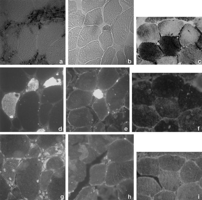
Figure 1.

Photomicrographs (×100) of serial sections from muscle biopsy of patient 1 (a, d, and g), patient 5 (b, e, and h), and normal control (c, f, and i). a to c: Histochemistry for glycogen phosphorylase; d to f: Immunohistochemistry for fetal myosin; g to i: Immunohistochemistry for muscle-specific glycogen phosphorylase.
