
Figure 2.
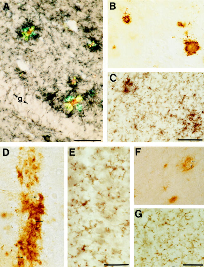
Activated microglia are associated with dense core and congophilic amyloid plaques but not with diffuse amyloid deposits. A: Double labeling of Mac-1 immunostaining (blue-black reaction product) and Congo red histochemistry revealed that virtually all birefringent plaques are surrounded by activated microglia. Shown is the molecular layer of the hippocampus of a 14 month-old APP23 transgenic mouse. g, granular cell layer. B and C: Similarly, serial sections alternately immunostained for Aβ and Mac-1 revealed that the vast majority of compact plaques were surrounded by intensely stained and hypertrophic Mac-1 positive microglia. D, E, F, and G: Occasionally, NT-11 positive diffuse amyloid was seen as a band in the neuron-rich layer II of the cingulate cortex (D) and in areas such as the striatum (F). Note that the diffuse amyloid spares cell bodies (arrows). No corresponding microglia reaction was observed in adjacent sections immunoreacted with Mac-1 (E and G). All calibration bars are 50 μm.