
Figure 5.
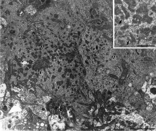
Electron micrograph of the interface between extracellular amyloid fibrils (A) and the cytoplasm of two microglial cells (M) in a 15-month-old APP23 transgenic mouse. Since a high concentration of glutaraldehyde was used in the fixative, the ultrastructure is better conserved compared to the immunoelectron micrographs shown in Figure 4, A and B ▶ . Within the microglia cytoplasm, pockets filled with parallel oriented amyloid fibrils (arrowheads) are observed. Cellular organelles such as rough endoplasmic reticulum (rer) and mitochondria (m) are in close vicinity of the amyloid fibrils. Insert: Amyloid bundles in the microglia cytoplasm surrounded by an incomplete membrane (arrows). Calibration bars, 1 μm and 0.5 μm (insert).