
Figure 6.
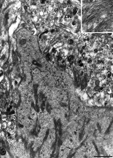
High-power electron micrograph of microglia cytoplasm with amyloid bundles (A) in a 15-month-old APP23 transgenic mouse. The cytoplasm reveals a dense-granular appearance which is rich in free polyribosomes (arrowhead) and rough endoplasmic reticulum (rer). Mitochondria (m) are also present. Occasionally a membrane surrounding the amyloid bundles is evident (arrow). Insert: Oriented amyloid fibrils in high magnification. DN, dystrophic neurite. Calibration bars, 1 μm and 0.2 μm (insert).