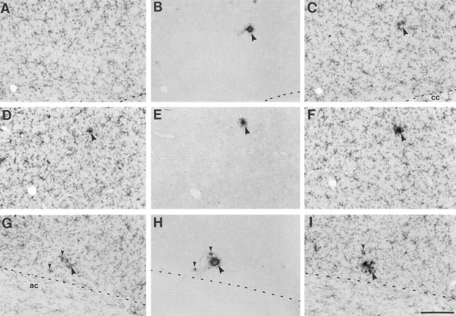
Figure 7.
Early amyloid microdeposits in young APP23 transgenic mice are of dense core nature and are surrounded by intensely Mac-1-immunostained microglia. Serial sagittal sections in young (4- to 9-month-old) transgenic mice were immunostained alternately for Mac-1 and Aβ, respectively. Three series of 3 serial sections are shown: A-C, parietal cortex layer V/VI; D-F, frontal cortex, layer III; G-I, ventral orbital cortex immunostained for Mac-1 (A, C, D, F, G, I) and Aβ (B, E, H). The amyloid deposits in each series are associated with a notable clustering of intensely stained Mac-1 immunoreactive microglia in either the preceding or the following section of the series. Note that the smallest amyloid plaques with a diameter of <5 μm were already dense-core in nature (small arrowheads in H) and associated with a few activated microglia. Abbreviations: cc, corpus callosum; ac, anterior commissure. Calibration bar, 100 μm (I). All prints have the same magnification.

