
Figure 1.
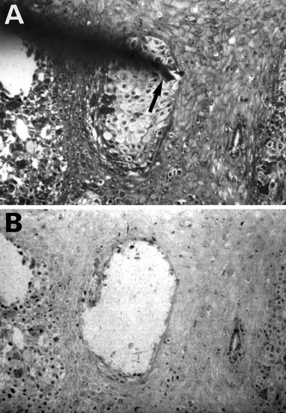
Microdissection of cutaneous MM. A: MM cells are arranged in irregularly shaped nests in the epidermis. The needle tip (arrow) is attached to a tumor cell nest. B: Tumor cell nest was dissected, leaving large holes behind. Original magnification, ×150