
Figure 3.
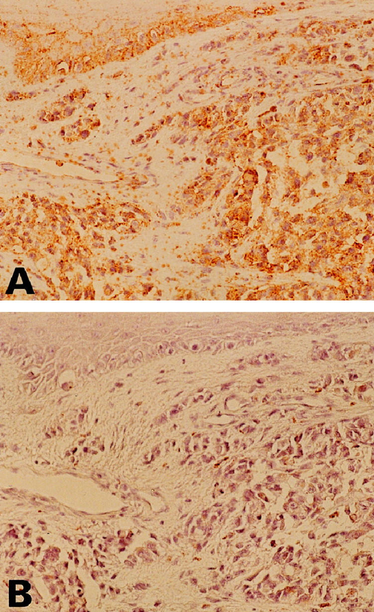
Visualization of Fas in cutaneous MM by immunohistochemistry. Antibodies were detected by a diaminobenzidine method that produces a brown color. Counterstaining of nuclei was with hematoxylin (blue). A: MM shows immunoreactivity for Fas. B: Negative control of Fas immunostaining. Fas antibody preincubation with the Fas peptide shows no detectable immunostaining in MM. (Original magnifications, ×100).