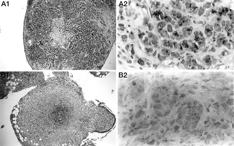
Figure 3.

Morphological study and immunohistological detection of TGF-β1 in tumor nodules from rats treated or not with the lipid A. Histological sections of tumor nodules were taken on day 28 after tumor cell injection from mesentery of untreated control rats (A) or after the fifth injection of OM 174 in treated animals (B). First, the sections in both groups were morphologically studied (A1, B1; HES: × 100). Then, the sections were stained with an anti-TGF-β1 antibody (A2, B2; AEC: × 1000).
