
Figure 1.
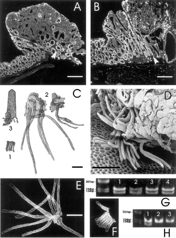
Demonstration of the presence of colossal crypts at the margin of colon adenomas from ApcMin mice and their Apc status. All scale bars represent 250 μm. A and B: Laser scanning confocal micrographs of frozen sections through colon adenomas from ApcMin mice. The sections have been stained with an antibody specific for the carboxy terminus of Apc. In A, note the absence of staining in the dysplastic tissue and the layer of Apc-positive crypts underlying the tumor (arrowhead). There are also some enlarged crypts, sectioned in a more longitudinal plane, at the left tumor margin. In B, note the large Apc-positive branching nondysplastic crypt at the tumor margin (arrowhead). C: Photomicrograph of intestinal epithelium isolated from an ApcMin mouse. A group of crypts from normal colon is labeled 1. Compare them with the two sets of colossal crypts from the margins of an adenoma, labeled 2. Note the branching colossal crypt on the right. A normal crypt-villus unit isolated from proximal jejunum, labeled 3, is shown for size comparison. D: Scanning electron micrograph showing the base of a large adenoma from the colon (note the honeycomb-like array of vacant pits in the lamina propria, from which crypts have been extracted by the isolation procedure). A few colossal crypts, which have been extracted from the margins of the tumor, remain attached to the tumor by surface epithelium. Many of the crypts are branched. The crypts shown are ∼500 μm long (approximately double the normal crypt height), although they appear smaller in this view due to foreshortening and partial obstruction of the crypt tops. Dysplastic epithelium is visible in the upper right corner. E and F: Fluorescence micrographs of isolated colossal (E) and normal crypts (F) stained for Apc protein with an antibody specific for the carboxy terminus. This demonstrates that the cells in colossal crypts are making full-length Apc. The figures are at the same magnification. G: Ethidium-bromide-stained gel showing PCR product from single colossal crypts (lanes 1 and 2) and groups of normal crypts (lanes 3 and 4). Note the presence of both wild-type (123-bp) and ApcMin (144-bp) alleles in both colossal and normal crypts. This confirms the antibody results presented in E and F. H: Demonstration of the loss of heterozygosity that occurs in dysplastic epithelium from ApcMin mice. Lane 1 shows PCR product from normal epithelium (with both Apc alleles) whereas lanes 2 and 3 show product from isolated dysplastic epithelium from adenomas (demonstrating only the ApcMin allele).