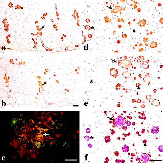
Figure 1.

Immunostaining with antibodies to Aβ and AMY 117 demonstrated the co-occurrence of the two antigens in the vast majority of plaques in AD brain. a and b: Adjacent 8-μm ethanol-fixed, paraffin sections of occipital cortex from a 90-year-old AD patient show overlapping immunoreactivities between Aβ labeled with R1282 (a) and AMY 117 (b); arrows mark one example. c: Double-immunofluorescent labeling with AMY 117 (ATC green) and the Ap antibody, Angela (Texas Red) on a 40-μm ethanol-fixed AD brain cryosection shows partial overlap in yellow (arrow) of the two antigens within an individual plaque lesion. The confocal image shown in c, kindly provided by Dr. M. L. Schmidt (The Center for Neurodegenerative Diseases, University of Pennsylvania School of Medicine, Philadelphia, PA), is the compilation of six images taken at 1-μm intervals. This image reflects the same yellow-labeled co-localization between AMY 117 and Aβ antibody R1282 we have observed in 8-μm AD brain sections by non-confocal fluorescent microscopy (data not shown). d to f: Three adjacent 8-μm briefly formalin-fixed, paraffin sections of frontal cortex from a 69-year-old AD patient illustrate overlapping Aβ and AMY 117 immunoreactivities. d shows plaques immunostained with Aβ antibody R1282, e shows plaques immunostained with AMY 117, and f shows double labeling with both antibodies (AMY 117 visualized in brown with DAB and Aβ (R1282) visualized in red with alkaline phosphate). The arrow indicates a large Aβ plaque surrounded by AMY 117 IR. The arrowheads indicate a single plaque that is negative for Aβ in the first section (d), shows AMY 117 IR in the second (e), and shows the presence of both antigens (red and brown reaction products) in the third section f.Note that the Aβ IR blood vessels (asterisk) shown in d and f are AMY 117 negative in e. Scale bars, 100 μm a, b, d to f and 10 μm (c).
