
Figure 3.
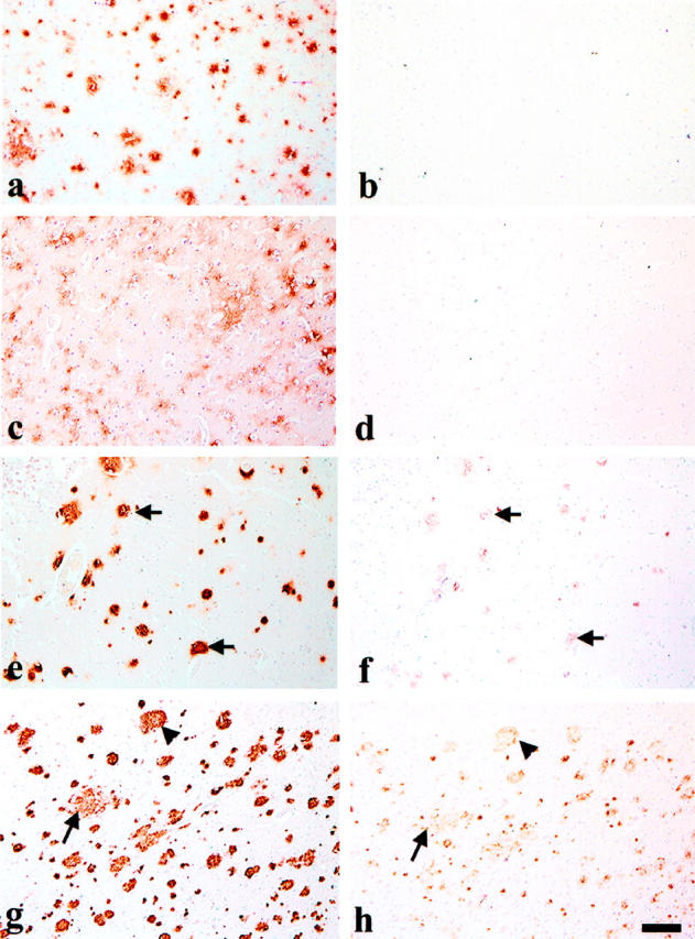
Aβ42 deposition precedes that of the AMY 117 antigen in young DS brain. Eight-micron, long-term formalin-fixed, paraffin sections from young DS brain were immunostained with Aβ42 antibody 21F12 and are shown in the left column (a, c, e, and g). Adjacent sections were immunostained with AMY 117 after double pretreatment of the tissue (microwaving and proteinase K) and are shown in the right column (b, d, f, and h). a and b: The frontal cortex of a 12-year-old DS patient shows many Aβ42 IR diffuse plaques (a) but no AMY 117 IR (b). c to f: Adjacent sections of a large block of tissue containing both temporal cortex and hippocampus from a 16-year-old DS patient show Aβ42 IR in many diffuse plaques in temporal cortex (c) and in more compacted plaques in hippocampus (e). AMY 117 IR is not seen in the temporal cortex (d) but is visible in some compacted plaques in the hippocampus (f). Arrows in e and f indicate examples of overlapping Aβ and AMY 117 immunoreactivities. g and h: Adjacent sections of frontal cortex from a 29-year-old DS patient show abundant IR for both Aβ (g) and AMY 117 (h). Arrowheads indicate relatively compacted plaques immunoreactive for both antigens, whereas arrows show a relatively less compated Aβ42 IR plaque that has less abundant, finely punctate AMY 117 IR. Scale bar, 100 μm for all images.