Figure 2.

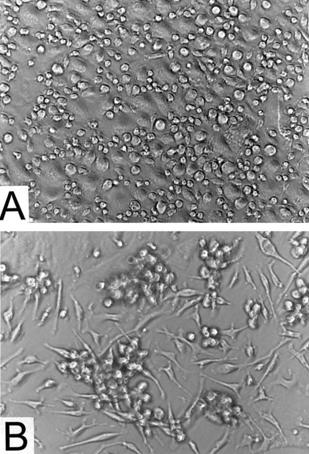
Cultures of adherent peritoneal cells viewed by phase-contrast microscopy. A: Cells maintained in the absence of rSAA2 and AEF. B: Cells incubated with rSAA2 and AEF; amyloid is seen at sites of clustered cells. Magnification, ×200.

Cultures of adherent peritoneal cells viewed by phase-contrast microscopy. A: Cells maintained in the absence of rSAA2 and AEF. B: Cells incubated with rSAA2 and AEF; amyloid is seen at sites of clustered cells. Magnification, ×200.